Introduction
Desmosomes, the junctions that hold both your skin and cardiac cells together, are said to be the strongest form of intercellular junctions in the human body. This strength is not without a reason, as the failure of desmosomes has been known to cause many cardiac diseases, and in some severe cases, cardiac arrest. The source of this strength was long debated, but was ultimately thought to stem from the adhesiveness and the rigidity of the desmosome structure, however, recent findings by a team of researchers at the University of Manchester suggest that the way in which desmosomes achieve this strength is actually due to flexibility and the extreme adhesion of the proteins these structures consist of.
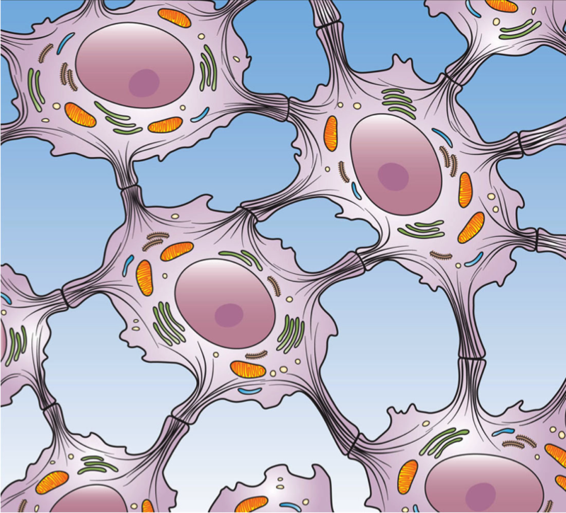
An artist’s rendition of desmosomes joining epithelial cells. CC BY 3.0 – https://en.wikipedia.org/wiki/Cell_junction#/media/File:Desmosome_Cell_Junction.png
Despite desmosomes being remarkably durable, mechanisms in which they can be damaged are not uncommon, nor are the diseases caused by the damaging of them. In some animals, infectious agents that release proteases or dermotases, enzymes that degrade the proteins in the bond between the desmosome and epithelial tissue have been observed and have been known to cause skin lesions and other skin conditions. Mutations in the genes of some proteins that compose desmosomal bonds have also been found to be the underlying causes of some heart diseases, such as arrhythmogenic right ventricular dysplasia, and with this discovery, the researchers hope to open up new directions in which research regarding these diseases can take place.
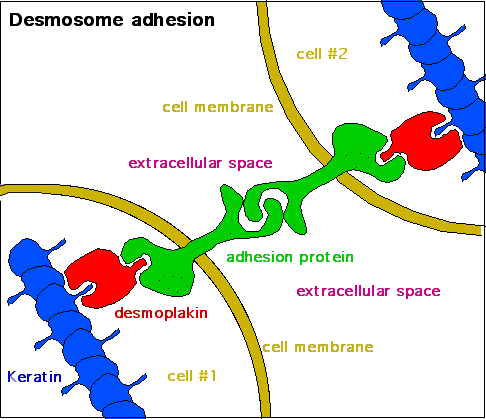
Diagram showing how the adhesion of desmosomes is achieved between two cells. CC BY-SA 3.0 – https://en.wikipedia.org/wiki/Desmosome#/media/File:Desmosome_-_2.png
Function and Form
Although the flexibility of desmosomal proteins has not been known of for a very long period of time, the strength in which desmosomal adhesion can be achieved has. This adhesion is maintained primarily by the individual strengths of the proteins that the desmosome is constructed of, which for the most part, is cadherins. Desmosomes are very dynamic in that they are not always extremely strong, and there are important physiological reasons for this. Desmosomes have been found to become weaker in periods of healing and development as this allows extracellular components that are associated with these processes to migrate with ease, ultimately making them more efficient.
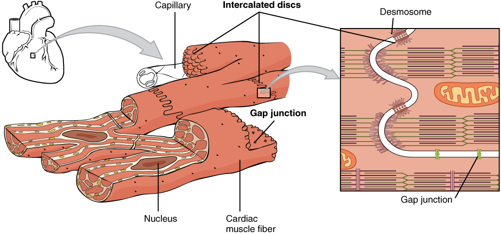
A diagram showing how cardiac muscle is connected. Both desmosomes and other forms of junctions such as gap junctions are utilized. CC BY 3.0 – https://en.wikipedia.org/wiki/Cardiac_muscle#/media/File:1020_Cardiac_Muscle.jpg
In the study conducted at the University of Manchester, the original objective was only to determine how desmosomes could achieve such great adhesion between cells. The discovery that the proteins linking cells together were flexible was shocking to Tabernero and her colleagues, and she described the findings as “the total opposite” of what was hypothesized. With this discovery, the researchers decided that testing some of the properties of these proteins could be beneficial. Tests were performed until a model could be constructed showing how the extracellular domains of the cadherin’s were much more ordered than that of other forms of cell junctions, something that the researchers believe to be the cause of the flexibility and strength of desmosomes.
Implications
The research team from the University of Manchester are hopeful that the discovery made will allow more research to be performed pertaining to cardiac diseases caused by the degradation of desmosomes, and that eventually treatments can be developed to aid patients who are either predisposed to such conditions, or even currently experiencing symptoms of them. Although research that is more specific to cardiac tissue disorders may not be completed for quite some time, it is still something to look forward to.
Recent Comments